Recombinant antibodies offer several key advantages compared to traditional antibodies. These include superior lot-to-lot consistency, continuous supply, and animal-free manufacturing. As such, recombinant antibodies are seeing increased use for scientific research, especially as a means of addressing the ongoing reproducibility crisis.
Traditional polyclonal and monoclonal antibodies are the product of normal B cell development and genetic recombination. They are generated by immunizing an animal with an antigen to elicit an immune response. While polyclonal antibodies are secreted by many different B cell clones and recognize multiple antigenic epitopes, monoclonals originate from a single B cell clone and are specific for just one epitope.
Recombinant antibodies are monoclonal, but their production involves in vitro genetic manipulation. After cloning the antibody genes into an expression vector, this is then transfected into an appropriate host cell line for antibody expression. Mammalian cell lines are most commonly used for recombinant antibody production, although cell lines of bacterial, yeast, or insect origin are also suitable.
Because recombinant antibody production involves sequencing the antibody light and heavy chains, it is a highly controlled and reliable process. In contrast, hybridoma-based systems for producing monoclonal antibodies are subject to genetic drift and instability, increasing the potential for lot-to-lot variability or loss of antibody expression. Recombinant antibodies are highly consistent from lot to lot, thereby ensuring reproducible experimental results.
In vitro methods for producing antibodies are amenable to large-scale production, meaning antibody availability is unlikely to become a limiting factor. Moreover, since the recombinant antibody sequence is known, continuity of supply is assured; in situations where an antibody will be used to support large, long-term studies, this can be an especially critical factor.
Unlike traditional methods for antibody production, recombinant approaches avoid the need to use animals. Where polyclonal antibodies are purified directly from the serum of the immunized host, and monoclonals are purified from either hybridoma-derived tissue culture supernatant or ascites, recombinant antibodies are instead purified from the tissue culture supernatants of transfected host cell lines. Regardless of whether an antibody is polyclonal, monoclonal or recombinant, it must always be properly validated in the intended application prior to experimental use. At CST, we adhere to the Hallmarks of Antibody Validation™, six complementary strategies for determining the specificity, sensitivity, and functionality of an antibody in any given assay. By carefully tailoring these strategies to each antibody product, we guarantee that CST antibodies will work as expected, to help you achieve results you can trust.

| Cat. # | Size | Price | Inventory |
|---|---|---|---|
| 13387S | 100 µl |
| REACTIVITY | H |
| SENSITIVITY | Endogenous |
| MW (kDa) | 150 |
| Source/Isotype | Rabbit IgG |
Product Information
| Application | Dilution |
|---|---|
| Western Blotting | 1:1000 |
For western blots, incubate membrane with diluted primary antibody in 5% w/v BSA, 1X TBS, 0.1% Tween® 20 at 4°C with gentle shaking, overnight.
NOTE: Please refer to primary antibody product webpage for recommended antibody dilution.
From sample preparation to detection, the reagents you need for your Western Blot are now in one convenient kit: #12957 Western Blotting Application Solutions Kit
NOTE: Prepare solutions with reverse osmosis deionized (RODI) or equivalent grade water.
Load 20 µl onto SDS-PAGE gel (10 cm x 10 cm).
NOTE: Loading of prestained molecular weight markers (#59329, 10 µl/lane) to verify electrotransfer and biotinylated protein ladder (#7727, 10 µl/lane) to determine molecular weights are recommended.
NOTE: Volumes are for 10 cm x 10 cm (100 cm2) of membrane; for different sized membranes, adjust volumes accordingly.
* Avoid repeated exposure to skin.
posted June 2005
revised June 2020
Protocol Id: 10
Human
Bovine, Dog, Horse
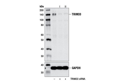
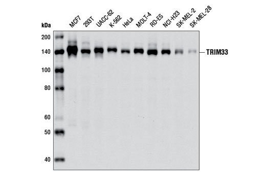
Monoclonal antibody is produced by immunizing animals with a synthetic peptide corresponding to residues near the carboxy terminus of human TRIM33 protein.
The transcriptional intermediary factor 1 (TIF1) family represents a group of proteins with multiple histone-binding domains. In humans, this family comprises four proteins, TIF1α/TRIM24, TIF1β/TRIM28/KAP1, TIF1γ/TRIM33/Ectodermin, and TIF1δ/TRIM66, which are characterized by an amino-terminal tripartite motif (TRIM) domain consisting of a RING domain, two B boxes, a coiled-coil domain, and a carboxy-terminal PHD finger and bromodomain (1). Despite their similar overall structure, these proteins have diverse roles in transcriptional regulation. TIF1α functions as a ligand-dependent nuclear receptor coregulator and more recently has been implicated in regulating p53 stability (2). TIF1β is an intrinsic component of the N-CoR1 corepressor complex and the NuRD nucleosome-remodeling complex (3) and functions as a corepressor for Kruppel-associated box (KRAB) zinc-finger transcription factors (4). Furthermore, TIF1β promotes heterochromatin-mediated gene silencing formation by serving as a cofactor for heterochromatin protein HP1 (5). TIF1δ expression is restricted to the testis and has been shown to interact with HP1γ (6).
In contrast, the ubiquitous nuclear protein TRIM33 does not interact with either HP1 family members or chromatin-remodeling/modifying complexes. Rather, TRIM33 plays a pivotal role in signaling cascades driven by the TGF-β superfamily of ligands (7-9). A research study suggests that TRIM33 and Smad4 compete for binding to receptor phosphorylated Smad2/3 and that TRIM33-Smad2/3 and Smad4-Smad2/3 complexes complement one another in the TGF-β-dependent control of hematopoietic cell fate (9). Other studies, however, demonstrate that TRIM33 functions to repress signal relay by the TGF-β superfamily (7-8,10). Indeed, knockout of murine Trim33 results in embryonic lethality due to upregulated Nodal signaling (10). Mechanistically, TRIM33 functions as an E3-ubiquitin ligase and promotes monoubiquitination of Smad4, a modification that impairs its ability to associate with phospho-Smad2 (8). This negative regulatory mechanism is further substantiated by the discovery that TRIM33 disrupts transcriptionally competent Smad complexes on the promoter/enhancer regions of TGF-β-responsive genes by associating with specific epigenetic marks on histone H3, which is a requirement for activating TRIM33's monoubiquitin ligase activity toward Smad4 (11). In line with the ability of TRIM33 to regulate the development of different blood cell lineages, it was shown that loss of TRIM33 expression due to epigenetic silencing of its promoter contributes to the pathogenesis of chronic myelomonocytic leukemia (12).
Except as otherwise expressly agreed in a writing signed by a legally authorized representative of CST, the following terms apply to Products provided by CST, its affiliates or its distributors. Any Customer's terms and conditions that are in addition to, or different from, those contained herein, unless separately accepted in writing by a legally authorized representative of CST, are rejected and are of no force or effect.
Products are labeled with For Research Use Only or a similar labeling statement and have not been approved, cleared, or licensed by the FDA or other regulatory foreign or domestic entity, for any purpose. Customer shall not use any Product for any diagnostic or therapeutic purpose, or otherwise in any manner that conflicts with its labeling statement. Products sold or licensed by CST are provided for Customer as the end-user and solely for research and development uses. Any use of Product for diagnostic, prophylactic or therapeutic purposes, or any purchase of Product for resale (alone or as a component) or other commercial purpose, requires a separate license from CST. Customer shall (a) not sell, license, loan, donate or otherwise transfer or make available any Product to any third party, whether alone or in combination with other materials, or use the Products to manufacture any commercial products, (b) not copy, modify, reverse engineer, decompile, disassemble or otherwise attempt to discover the underlying structure or technology of the Products, or use the Products for the purpose of developing any products or services that would compete with CST products or services, (c) not alter or remove from the Products any trademarks, trade names, logos, patent or copyright notices or markings, (d) use the Products solely in accordance with CST Product Terms of Sale and any applicable documentation, and (e) comply with any license, terms of service or similar agreement with respect to any third party products or services used by Customer in connection with the Products.